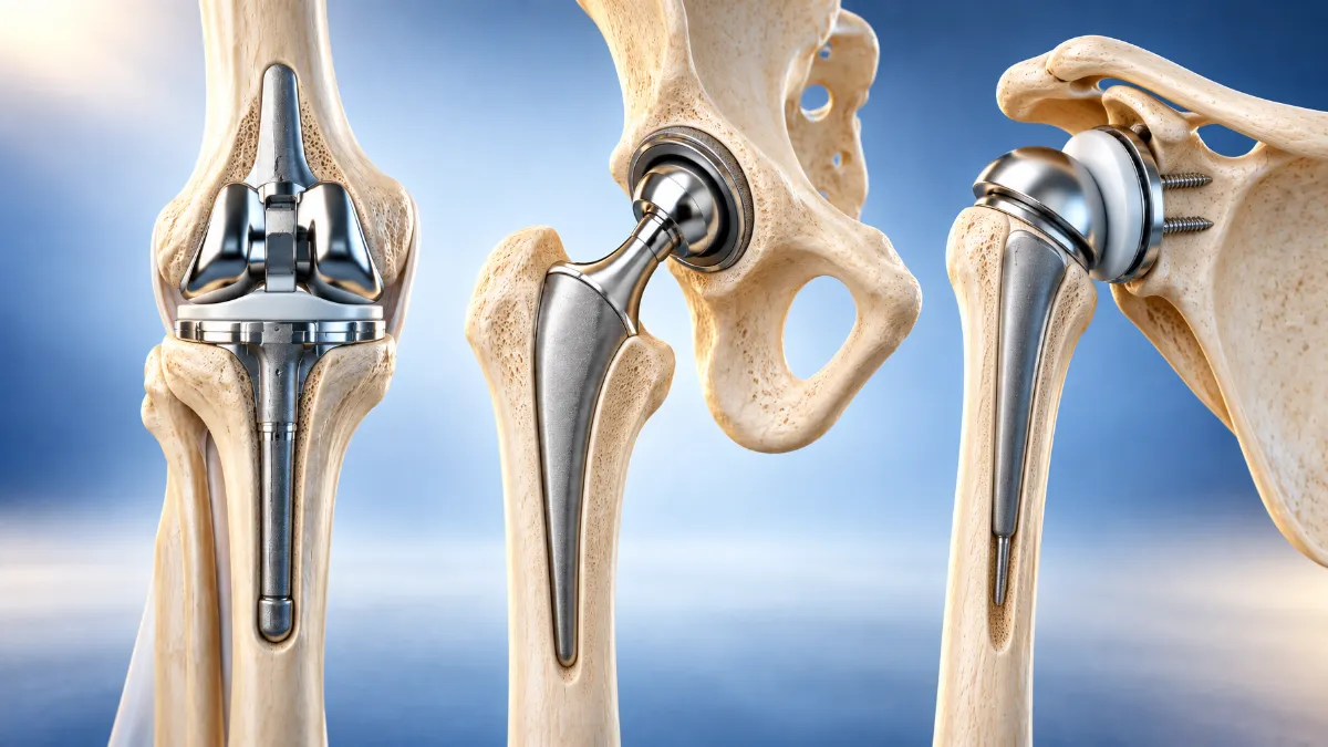
Surgical and non-surgical care for joints, ligaments, fractures and knee alignment — tailored to your diagnosis and goals. Consultations at Suyash Hospital and Balaji Medicare, Raipur, and the visiting clinic in Bilaspur.
Joint Replacement
Knee, hip & shoulder replacement surgery
Joint replacement (arthroplasty) replaces a worn or damaged joint surface with an implant made of metal, high-grade plastic or ceramic. It is usually considered when arthritis or an old injury causes persistent pain and stiffness that limits walking, sleep or daily activity, and non-surgical care — medicines, physiotherapy, activity changes, injections — is no longer enough.
Dr. Mittal evaluates each case individually, including X-rays and limb alignment, to decide whether replacement is appropriate and which type suits you.
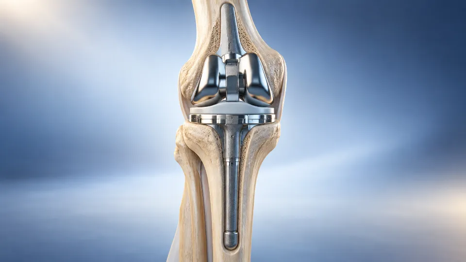
Knee, hip and shoulder replacement implants (illustration)
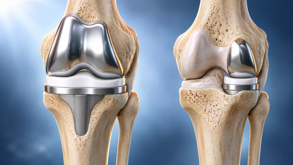
Knee replacement (total & partial)
The worn surfaces at the end of the thigh bone (femur) and shin bone (tibia) are replaced with a metal and plastic implant. When arthritis is confined to one part of the knee, a partial (unicondylar) replacement may be an option.
Commonly considered for: advanced osteoarthritis, rheumatoid arthritis, arthritis after injury, and bow-leg or knock-knee deformity caused by arthritis.
Procedure & recovery
Surgery is usually done under spinal or regional anaesthesia. Walking with support generally begins within a day or two, and hospital stay is commonly around 3–4 days. Physiotherapy then focuses on bending, straightening and strengthening the knee, progressing from a walker to a stick over the following weeks. Individual timelines vary.
Possible risks — including infection, blood clots, stiffness and implant wear over time — are discussed in detail before surgery.
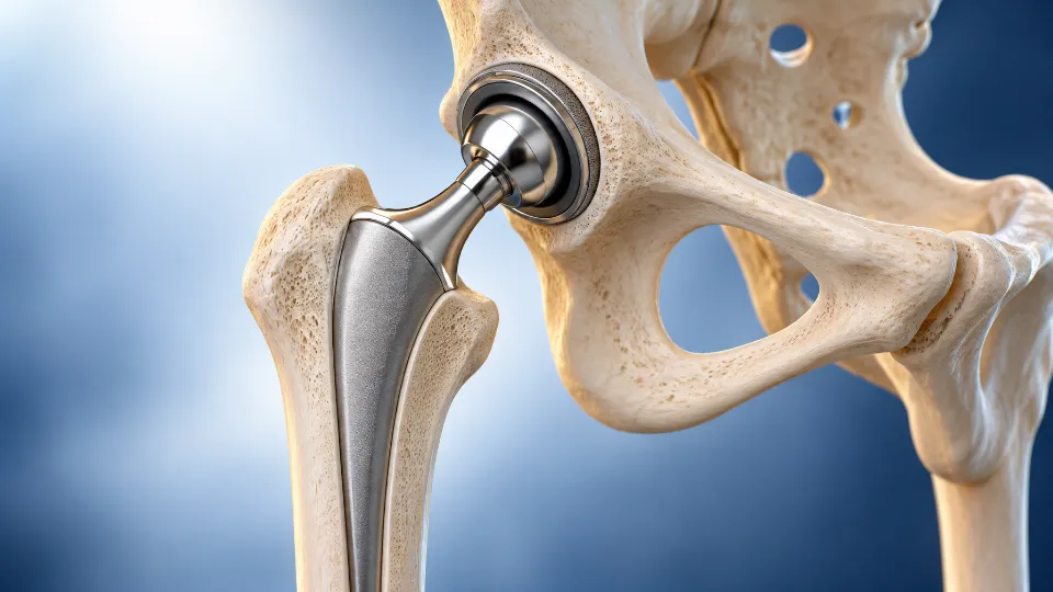
The damaged ball (femoral head) and socket (acetabulum) of the hip are replaced with an implant, relieving pain from a worn joint and helping restore movement.
Commonly considered for: hip osteoarthritis, avascular necrosis (AVN) of the femoral head, inflammatory arthritis, arthritis after old injuries, and some hip fractures in older patients.
Procedure & recovery
Standing and walking with support usually start soon after surgery. Physiotherapy and simple precautions guide the first few weeks while the tissues around the new joint heal. Recovery pace differs from person to person.
Risks such as infection, blood clots, dislocation and a difference in leg length are explained and planned for before surgery.
Total shoulder replacement
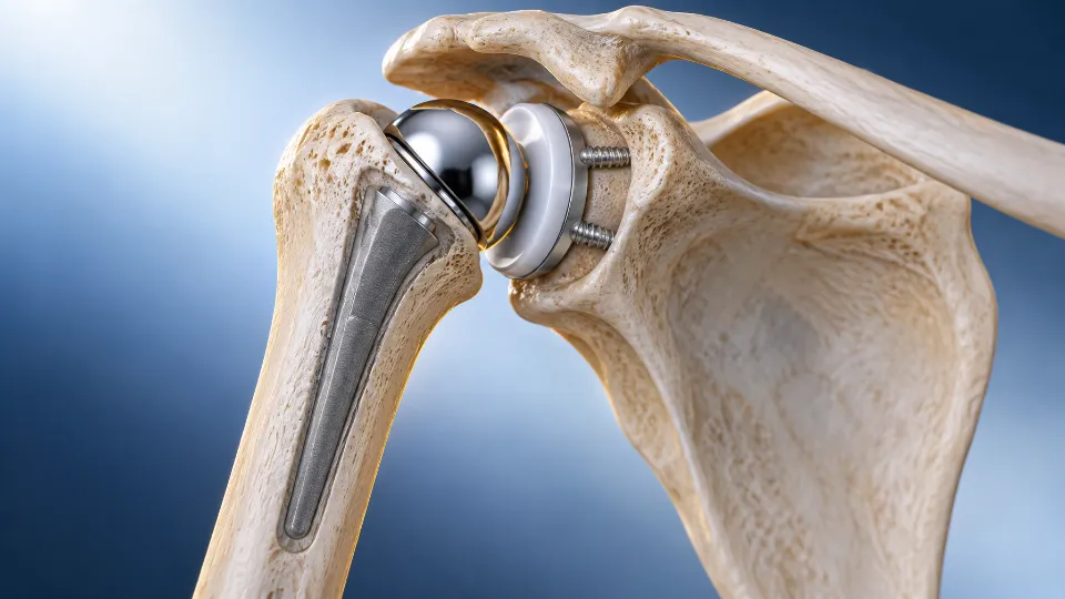
The worn ball (humeral head) and socket (glenoid) of the shoulder are replaced with an implant. Where the rotator cuff is torn beyond repair, a reverse shoulder replacement — which swaps the positions of the ball and socket — may be advised.
Commonly considered for: shoulder arthritis, rotator cuff tear arthropathy, avascular necrosis, and some complex upper-arm fractures in older patients.
Procedure & recovery
The arm is supported in a sling for the first few weeks, followed by staged physiotherapy — gentle movement first, then strengthening. Recovery of comfortable daily use typically unfolds over several months and varies between individuals.
A repeat operation to replace part or all of an existing knee or hip replacement that has become loose, worn, unstable or infected.
Commonly considered for: new or increasing pain in a replaced joint, instability, implant loosening or wear, infection, or a fracture around the implant.
Procedure & recovery
Revision surgery is more involved than a first replacement. Detailed imaging and blood tests help identify the cause, and the plan — including implants and any bone grafting — is tailored to each case. Rehabilitation is usually more gradual.
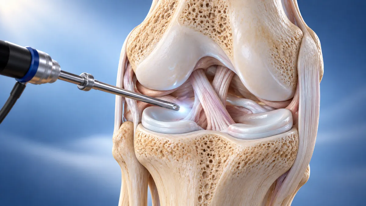
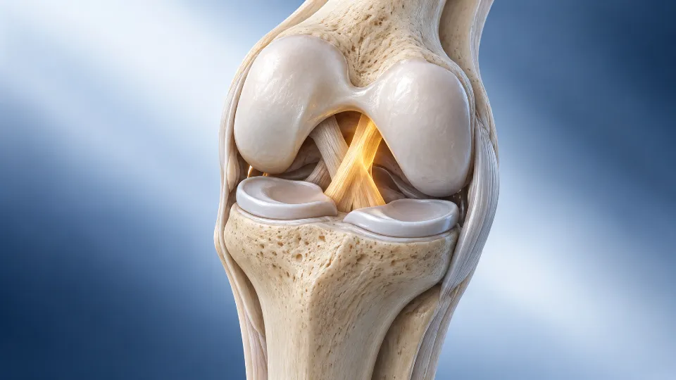
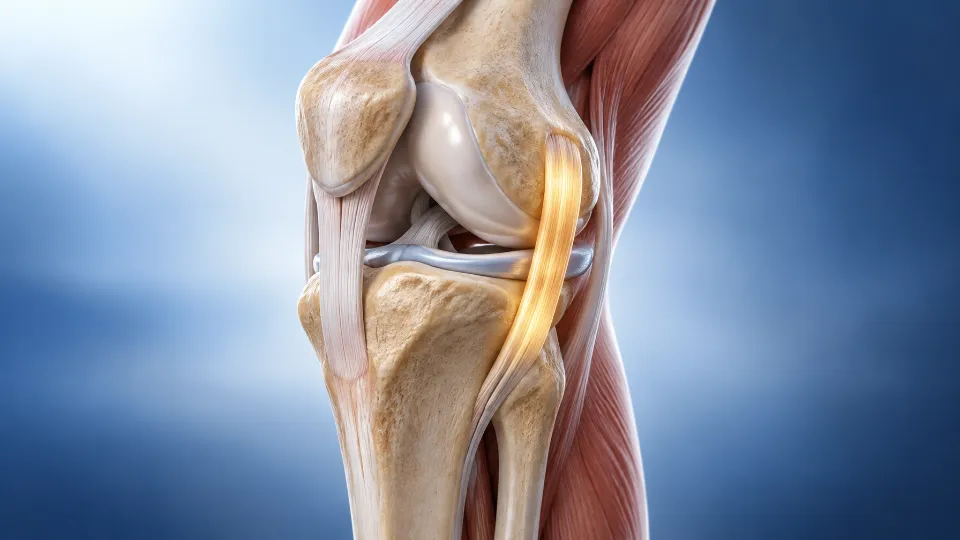
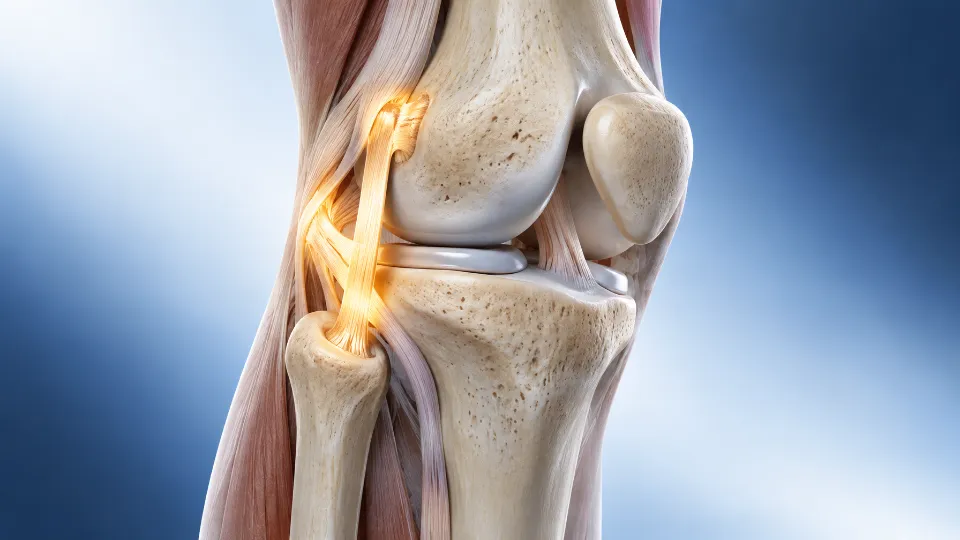
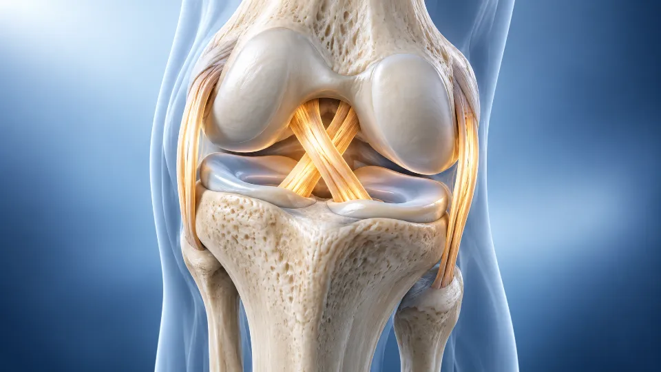
Knee joint ligaments and menisci with an arthroscope (illustration)
Knee Arthroscopy & Ligaments
Keyhole knee surgery and ligament reconstruction
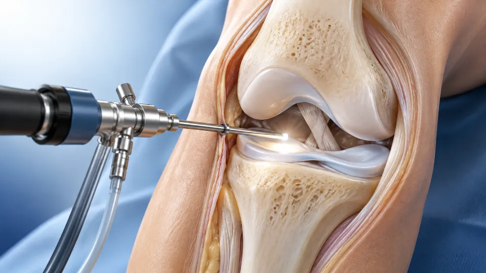
Knee arthroscopy uses a small camera and fine instruments through keyhole incisions to diagnose and treat problems inside the knee, typically allowing a faster recovery than open surgery.
Ligament injuries — from sport, falls or road accidents — are among the most common causes of a knee that feels unstable or gives way. Treatment ranges from bracing and physiotherapy to arthroscopic reconstruction, depending on which ligaments are injured and how you use your knee.
Repair or trimming of meniscus tears, treatment of cartilage (chondral) injuries, removal of loose bodies, and diagnostic arthroscopy for unexplained knee pain or locking.
Commonly considered for: pain, swelling, catching or locking after a twisting injury, with matching MRI findings.
Procedure & recovery
Many procedures need only a short hospital stay. After a meniscus repair, weight-bearing and bending may be limited for some weeks to protect the stitches; after trimming, recovery is usually quicker.
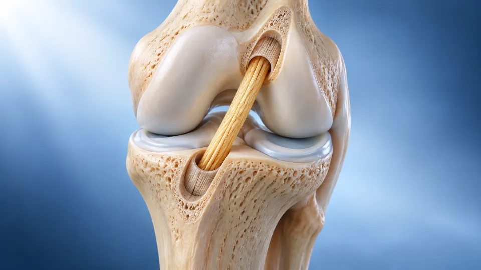
The anterior cruciate ligament (ACL) controls forward movement and rotation of the shin bone. A torn ACL can make the knee give way when turning or pivoting. Reconstruction replaces it with a graft — usually your own tendon — through keyhole surgery.
Commonly considered for: knee instability after an ACL tear, active people and athletes, and ACL tears with associated meniscus or cartilage damage.
Procedure & recovery
Graft choice is tailored to your activity level. A structured rehabilitation programme follows, and return to sport is commonly discussed in the range of about 6–9 months — general information, not a personal prognosis.
The posterior cruciate ligament (PCL) stops the shin bone sliding backwards. PCL tears often follow a direct blow to the front of the knee — such as a dashboard injury in a road accident — or a fall on a bent knee.
Commonly considered for: back-of-knee pain and instability after such injuries, and PCL tears combined with other ligament damage.
Treatment options
Many isolated PCL injuries are managed with a brace and focused physiotherapy. Complete tears with ongoing instability, bony avulsions, or PCL injuries combined with other ligament tears may need arthroscopic fixation or reconstruction.
MCL injury treatment
The medial collateral ligament (MCL) runs along the inner side of the knee and resists knock-knee forces. It is commonly injured by a blow to the outer side of the knee or a twisting fall.
Commonly considered for: inner-knee pain, swelling and a feeling of the knee opening up on the inside.
Treatment options
Most MCL injuries heal well with a hinged knee brace and physiotherapy. Surgical repair or reconstruction is considered for severe tears, persistent instability, or when the MCL is torn together with the ACL or other ligaments.
LCL & posterolateral corner injury
The lateral collateral ligament (LCL) on the outer side of the knee is often injured together with neighbouring structures, collectively called the posterolateral corner.
Commonly considered for: outer-knee pain and instability after a blow to the inner side of the knee or a hyperextension injury.
Treatment options
Mild, isolated injuries may settle with bracing and rehabilitation. Complete tears — especially with posterolateral corner or cruciate ligament damage — often need surgical repair or reconstruction, ideally planned early. The nearby peroneal nerve is checked as part of the assessment.
Multiligament knee injury
Injury to two or more major knee ligaments, usually after high-energy trauma such as a road accident, and sometimes with a knee dislocation.
Commonly considered for: severe knee injuries with gross instability, often alongside other injuries.
Treatment options
These injuries need prompt assessment of the blood vessels and nerves around the knee. Reconstruction of the torn ligaments is planned in one or more stages, followed by a longer, carefully supervised rehabilitation.
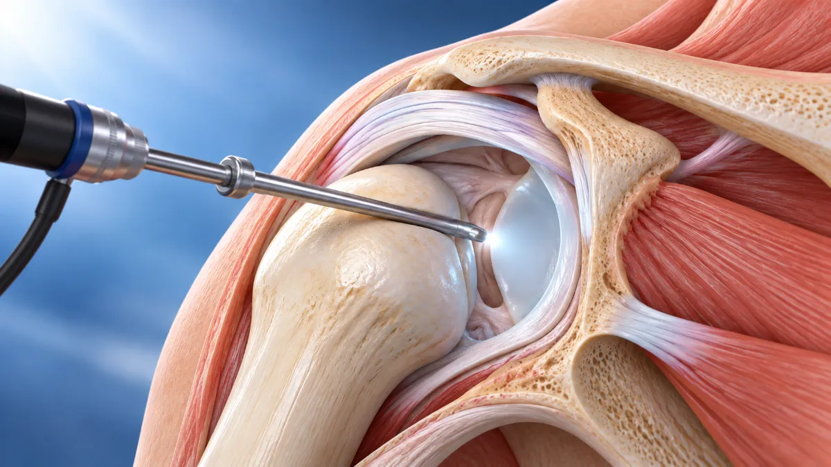
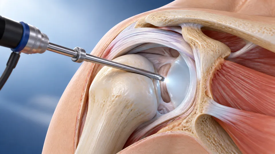
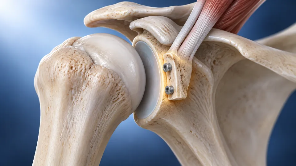
Shoulder arthroscopy treats pain, weakness and instability from rotator cuff tears, impingement and recurrent dislocation through small portals rather than an open incision.
When a shoulder keeps dislocating, the right operation depends on the damage to the labrum and to the bone of the socket and ball — assessed carefully on MRI and, where needed, CT scan.
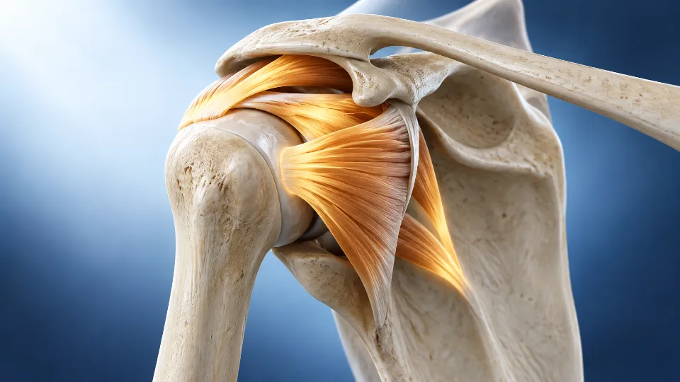
Shoulder joint with rotator cuff and labrum, and an arthroscope (illustration)
Shoulder arthroscopy
Keyhole surgery for impingement (subacromial decompression), biceps and labral (SLAP) problems, frozen shoulder release where indicated, and diagnostic arthroscopy.
Commonly considered for: shoulder pain or stiffness that has not improved with physiotherapy, medicines or injections.
Procedure & recovery
Most procedures involve a short hospital stay. A sling may be used for comfort, and physiotherapy starts early to regain movement. Recovery depends on the procedure performed.
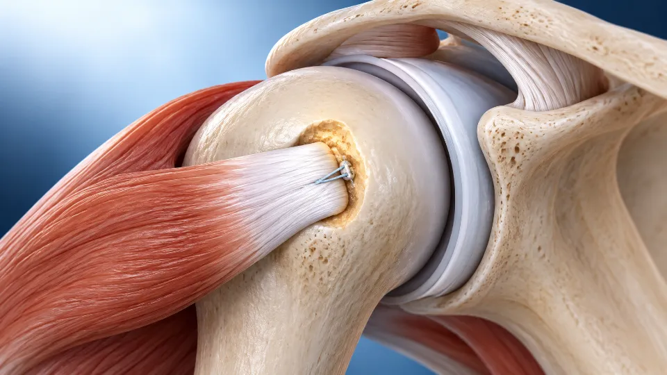
The rotator cuff is a group of four tendons that lift and rotate the arm. Tears can follow an injury or develop with age and wear, causing night pain and weakness when raising the arm.
Commonly considered for: full-thickness tears, tears after injury, and persistent pain and weakness despite physiotherapy.
Procedure & recovery
Arthroscopic repair reattaches the torn tendon to bone using small suture anchors. The arm is protected in a sling for several weeks while the tendon heals, followed by staged physiotherapy over the next few months.
Bankart repair
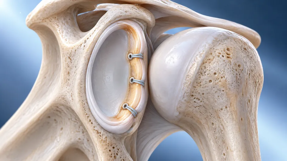
After a shoulder dislocation, the labrum — the cartilage rim of the socket — can tear away from the bone (a Bankart lesion), leaving the shoulder prone to dislocating again. Arthroscopic Bankart repair reattaches it using suture anchors.
Commonly considered for: recurrent dislocation or a shoulder that feels unstable, particularly in young and active patients.
Procedure & recovery
The shoulder is rested in a sling for the first few weeks, then movement and strength are rebuilt gradually. Return to contact sport is planned with your surgeon and physiotherapist.
Latarjet procedure
A small piece of bone (the coracoid process), with its attached tendons, is transferred to the front of the shoulder socket to restore lost bone and add stability.
Commonly considered for: recurrent dislocation with significant bone loss from the socket, or instability after a previous stabilisation operation.
Procedure & recovery
Planning is based on CT measurement of bone loss. A period in a sling is followed by progressive physiotherapy; the transferred bone takes a few months to heal fully.
Remplissage
Repeated dislocations can leave a dent in the back of the ball of the shoulder (a Hill–Sachs lesion). Remplissage fills this defect by fixing part of the rotator cuff tendon into it, so it no longer catches on the edge of the socket.
Commonly considered for: recurrent instability with a significant Hill–Sachs lesion — usually performed together with a Bankart repair.
Procedure & recovery
Done arthroscopically alongside the labral repair, with a similar sling period and staged rehabilitation.
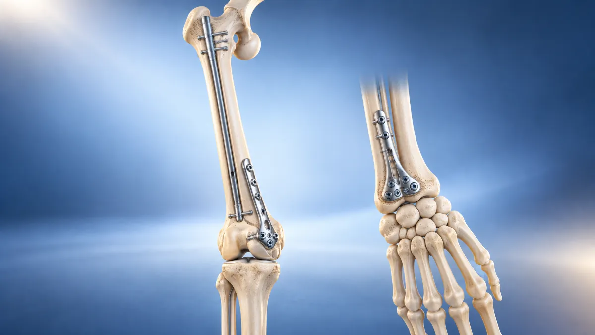
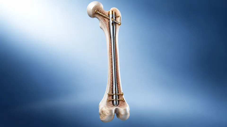
Fracture fixation with a femoral nail and bone plates (illustration)
Trauma & Fractures
Fracture and complex injury care
Fractures from road accidents, falls and sport need careful assessment — especially when they involve a joint, the pelvis, or several bones at once.
The aim of treatment is to restore alignment and stability so that movement can begin as early as it is safe, reducing the risk of long-term stiffness and arthritis.
Severe limb injuries involving multiple fractures, open (compound) fractures, or fractures with significant soft-tissue damage — often from road traffic accidents or falls from a height.
Treatment may include: open reduction and internal fixation (ORIF) with plates and screws, intramedullary nailing, external fixation for unstable injuries, and bone grafting.
What to expect
Treatment is often staged — initial stabilisation and wound care first, then definitive fixation once the soft tissues allow. Recovery ranges from several weeks to a few months depending on the injury, with a rehabilitation plan at every stage.
Periarticular fractures
Fractures near or into a joint — such as around the knee (tibial plateau, distal femur), ankle, elbow, wrist or shoulder. Because the joint surface is involved, accurate realignment matters.
Commonly presents with: severe pain, swelling and bruising around a joint, deformity, and difficulty moving the joint or bearing weight.
Diagnosis & treatment
X-rays, and often a CT scan, show the fracture pattern. Stable fractures may be treated in a cast or brace; displaced fractures usually need ORIF to restore the joint surface, followed by early, guided movement to limit stiffness.
Pelvic & acetabular fractures
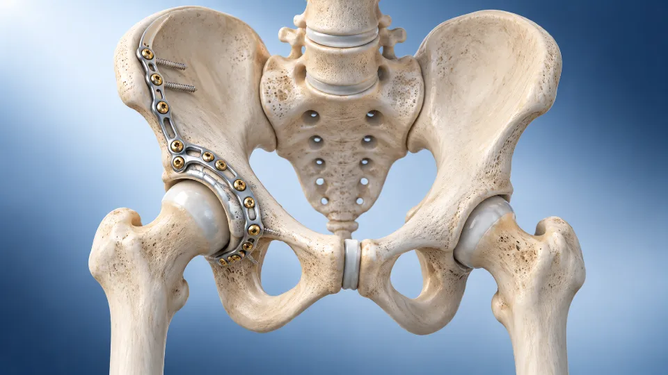
Fractures of the pelvic ring or the hip socket (acetabulum), usually from high-energy injuries. These can be associated with blood loss and other injuries, so emergency care comes first.
Commonly presents with: severe hip or groin pain, swelling, and inability to bear weight on the affected leg.
Diagnosis & treatment
CT-based planning guides treatment. Displaced fractures are usually fixed surgically with plates and screws to restore the socket and pelvic ring, followed by a period of protected weight-bearing and rehabilitation.
Knee Realignment (Osteotomy)
Joint-preserving knee realignment
Some knee problems are best treated by correcting the alignment of the bone itself. An osteotomy shifts load away from a worn part of the knee, which can help younger, active patients keep their own joint and delay or avoid the need for a replacement.
High tibial osteotomy (HTO)
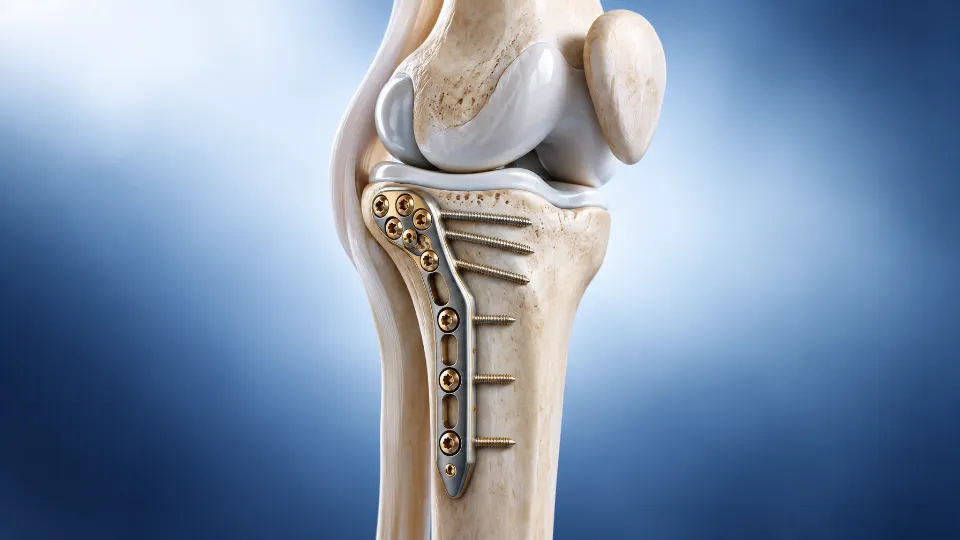
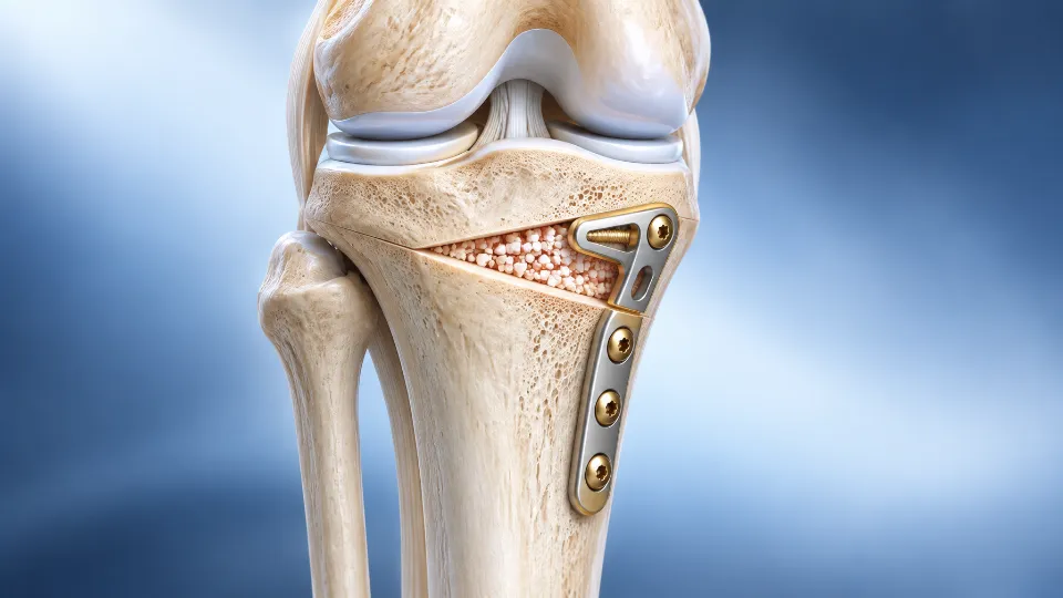
The shin bone is realigned just below the knee to shift weight away from a worn inner (medial) compartment — most often in bow-legged knees with early arthritis — while preserving your own joint.
Commonly considered for: younger, active patients with medial knee arthritis and bow-leg (varus) alignment; sometimes combined with cartilage or ligament procedures.
Procedure & recovery
The bone is held with a plate while it heals. Weight-bearing is usually protected for some weeks, and bone healing typically takes a few months.
Distal femoral osteotomy (DFO)
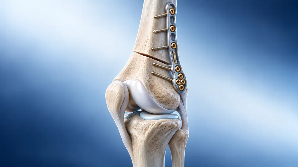
The lower thigh bone is realigned just above the knee — most often for knock-knee (valgus) alignment with wear in the outer (lateral) compartment, and in selected cases of kneecap maltracking.
Commonly considered for: younger patients with lateral knee arthritis and valgus alignment who wish to preserve their natural knee.
Procedure & recovery
Fixed with a plate, followed by protected weight-bearing and physiotherapy while the bone unites over a few months.
What To Expect
Your treatment journey
1. Consultation
A detailed history, examination, and review of any existing scans or reports to reach an accurate diagnosis.
2. Treatment planning
A discussion of all appropriate options — surgical and non-surgical — along with expected outcomes and risks.
3. Surgery (if needed)
Procedures are planned and explained in advance, including anaesthesia, hospital stay, and precautions.
4. Recovery & rehab
Structured follow-up and rehabilitation guidance to support a safe return to daily life or sport.
Not sure which treatment is right for you?
Book a consultation and get a clear, personalised recommendation based on your history and imaging.